Glossary
Unwanted voltage fluctuations due to the ECG signal, typically present when EMG electrodes are applied to the chest, back or abdominal muscles. ECG interference can be attenuated by processing the EMG signal post-recording, as it may not be sufficiently reduced by differential detection or the DRL technique.
(McManus et al., 2021)Sensor made of conductive material that couples the electrical potentials generated within the body to an electronic device. In EMG, reference electrodes are typically placed over an electrically silent area such as a tendon or bone (surface electrodes) or on the cannula (intramuscular Electromyographic signal electrode). In monopolar recording configurations, the detected EMG signal is the potential difference between the recording electrode and the reference electrode.
(McManus et al., 2021)The opposition (impedance) to the flow of the electrical current, as a function of frequency, at the contact point between the electrode and the skin surface. Lower electrode impedance is generally desired.
(McManus et al., 2021)The contact region between the electrode and the skin surface.The change in the current carrier (from ions to electrons) at the interface between the electrode and the skin generates random noise, the amplitude of which will depend on the contact surface, the electrode used and the skin condition. The impedance at the electrode-tissue interface may be reduced by conductive gel.
(McManus et al., 2021)The time-varying electrical potential recorded within a muscle, in the surrounding tissue or at the skin surface during muscle activity. It is the summation of the extracellular action potentials generated by muscle fibers within the vicinity of the electrode.
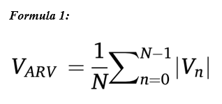
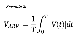
(McManus et al., 2021)An estimate of the average amplitude of a signal over a given time period. The ARV is defined as the average of the “rectified” (absolute value) signal over an epoch (or window) of duration T (or N samples). The average rectified value for one signal epoch, VARV, is defined as in Formula 1. N is the total number of samples being averaged (over the epoch of the EMG signal under analysis) and Vn is the value of the nth sample within the epoch. The epoch/window duration must be stated when reporting the VRMS. In practice, the mean value of the signal should be subtracted from the total signal length before rectifying. The epoch/window duration should also be stated when reporting VARV. The equation for VARV can also be expressed in continuous time, as shown in Formula 2. T is the length of the signal epoch in seconds.
(McManus et al., 2021)

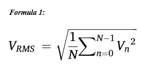
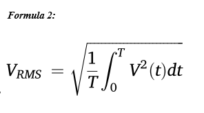
An estimate of the standard deviation (amplitude) of the signal over a given time window or epoch. The RMS value is the square root of the mean square value estimated over an epoch (or window) of duration T (or N samples). RMS is the square root of the power of the signal. The root mean square value, VRMS, for one signal epoch is defined in Formula 1, where N is the total number of samples being averaged (over the epoch of the EMG signal under analysis) and Vn is the value of the nth sample within the epoch. The epoch/window duration must be stated when reporting the VRMS. The equation for VRMS can also be expressed in continuous time as expressed in Formula 2, where T is the length of the signal epoch in seconds.
(McManus et al., 2021)

The instantaneous amplitude of the EMG signal is a measure of how far, and in what direction, the signal differs from zero Volts.The instantaneous amplitude of a signal differs from the magnitude as it can be either positive or negative. It is not recommended as an amplitude indicator for interference EMG signals.
(McManus et al., 2021)The peak-to-peak amplitude refers to the difference between the maximum and minimum values of the recorded waveform observed in a specified time window (epoch). Can be used to quantify the amplitude of motor unit action potentials and compound muscle action potentials. It is not recommended to use peak-to-peak amplitude as an amplitude indicator for the interference EMG signals that arise from the asynchronous firing of motor units during voluntary contractions.
(McManus et al., 2021)A smooth curve that tracks changes in the amplitude of an EMG signal over time.The envelope of an EMG signal is usually estimated by rectifying and then low-pass filtering the EMG signal. When reporting the EMG envelope, the filter characteristics (filter type, order, and cut-off frequency) or duration (and overlap) of the moving average window should be specified.
(McManus et al., 2021)We value your feedback
Let us know how helpful you found the recommendations above and how we can improve: